Thursday, January 27, 2011

Cosmetic surgeons paying marketing companies for positive comments and top ratings

I wasn't planning on writing this post, but after coming across some information today, I felt I had a  moral duty to report this. Some of you may be aware of this already. But for those consumers who are in the market looking for a cosmetic surgeon, this should be very enlightening.

Many people today use the Internet as tool for searching out the best cosmetic surgeon. You will find many doctor rating sites, and also websites rating the cosmetic surgeons website. But do you know how the surgeons sites are rated??? By what scientific study??? By what public voting system??? Well one of them that I had looked at to register my blogs site, gave me options in different purchases. The more you pay the better and higher on the list your website will be rated. Wow!!! I chose not to complete my registering. But many recognizable cosmetic surgeons are on that site marketing themselves. I have nothing against surgeons marketing themselves, but I do have a problem with misleading advertising.

On some doctor ratings websites,  former unhappy patients have accused some posters as being impostors, giving raving reviews to a particular surgeon. They believe the raving reviews are posted by the surgeons office staff, or possibly the surgeon themselves. On flip side it is possible that some of the negative posts are posted by people that aren't even former patients. That's a whole other  issue I will discuss in another post.

However would you believe that some of the glowing posts on  message boards and rating sites are posted by professional marketing companies hired by cosmetic surgeons? Here's some information from the blog of deceased plastic surgeon, Dr. Frank Ryan.

"I recently met the CEO of a web marketing company who described a type of marketing that his company does in which he and his team get paid by doctors to regularly log onto all the plastic surgery message boards and rave about those doctors. The CEO’s employees pose as patients and rave about what a wonderful surgeon so-and-so is and how they wouldn’t go anywhere else and how the doctor changed their lives, etc. The more the doctor pays, the more time the team will spend posting glowing reviews about the doctor. The CEO said", “If you visit some of these web sites, you will see names of doctors you never heard of with rave reviews and ten stars out of ten. You will also notice that some of the top doctors have fewer rave reviews, fewer stars and often more negative comments.”

This is where I feel the line has been crossed between ethical and unethical marketing. There are as some of you may already know, some cosmetic surgeons who are demanding rating doctor sites to remove negative comments and asking new patients to sign 5 year gag orders. It would be hypocritical for any of those doctors to do so, if they are involved in any deceptive marketing strategy's mentioned above. Even legally they would probably have a hard time suing a poster for negative comments that aren't warranted,  if they themselves are engaging in such deception. Hopefully there aren't too many cosmetic surgeons involved in such practices. So next time you see a cosmetic surgeon with all high marks, and raving reviews, you may want to take it with a grain of salt.

One of the best methods for finding a credible and competent surgeon is to get a referral from another surgeon or doctor.  If you can't get one in your city, then one site that you can is called Realself.com They are cosmetic surgeons who answer your questions, and will recommend a surgeon in a certain region or State. Another thing you can do to protect yourself somewhat is when your on message boards/forums is ask another poster who has commented on a surgeon a lot of questions about the surgery, facility, office staff, etc.. and you can ask them for before and after pictures of themselves (via private message or public). But keep in mind anyone can pull before and after pictures off the internet and claim its them or even photo shop pictures. You could ask them if they have family photo's as well and mention that they can block out their eye section if they like. Even better, is to use Skype, where you can see each other live via video camera. However don't get or use a real cheap camera, you may have troubles with motion. You don't need a very expensive one either, just very average. One site that allows you to interact with other posters is makemeheal.com  within their message boards section.

Some surgeons may even have great b/a pictures, and that's great assuming it's their own clients, but if you have a breathing issue, you need more reassurance. My last surgeon who was in his fifth year, had marketed himself very well on his website, youtube, and in cosmetic magazines. He convinced me that he performed more nose surgery's then the average surgeon. So I assumed his 5 years may be equal to 10 years of another surgeon. Big mistake. I ended up much worse off as I have already mentioned on other posts. This doesn't mean a young surgeon can't be a good choice. If you can't afford or for whatever reason don't feel as comfortable with an experienced surgeon, then seeking out a younger surgeon who specializes in nose surgery makes sense in some cases. Just ask a lot of questions and see if your comfortable with the answers. Don't feel pressured or desperate to give in to what ever they say. That's a red flag. Find out who the surgeon trained with? Did he train with any well known revision nose surgeon? Did he go beyond his regular specialty board license, for extra training in facial cosmetics? Did he graduate at top of his class? Did he go to reputable medical college? Has he conducted any research on the subject? Does he perform all different types of nose grafts for improving and strengthening the structure of the nose or does he just offer risky reductive surgery? Does he harvest rib, septal, and ear cartilage in any of his surgery's? Does his website dedicate a lot of information to nose revision surgery? Does the website indicate that he is knowledgeable in regards to the complexity and major issue's with correcting failed operated noses? Does he work in an office with other more experienced surgeons or does he work alone? 

http://www.dr-frank-ryan.blogspot.com/2009/12/truth-about-message-boards-and-plastic.html

Here's a site that specializes in Marketing to cosmetic surgeons.  I'm only including it here to show that such a company exists. I am not suggesting in any way that they engage in any of the above questionable practices.
http://www.surgeonsadvisor.com/

Wednesday, January 19, 2011

The Nose Tip: Dimensional Analysis 101

Advanced Therapy in Facial Plastics and Reconstructive Surgery  By: Dr. Regan Thomas

Tip Support Mechanisms
 Nasal tip shape is largely determined by the shape and integrity of the LLC (lower lateral cartilage). The tip surface is divided into the dome, alae, soft triangles, and columella. The major tip support mechanisms include the size, shape and integrity of the LLC, the attachment of the medial crus to the septal cartilage, and the attachment of the LLC to the ULC (upper lateral cartilage).  The minor support mechanisms are the interdomal ligament, dorsal portion of anterior septal angle, sesamoid cartilages, the LLC attachment to skin/soft tissue, and the nasal spine.


 http://www.lamfacialplastics.com/resources/learning-modules/rhinoplasty-tutorial/

Nasal Profile Projection   Minas Constantinides M.D. and Michael Carron M.D.
Unlike nasal length, which is relatively difficult to change, nasal projection can be changed easily. Because of this ease, however, changes are often overdone. Secondary corrections of overdone projection changes are among the biggest challenges faced in revision rhinoplasty.  Projection of the tip is the actual measured distance from the alar-facial plane to the tip. A change in nasal projection requires either an increase or decrease in the distance the nasal tip extends from the vertical facial plane. The Goode method and the 3:4:5 triangle are the two most common ways of measuring projection. [See Fig.23-4, Fig 23-5]

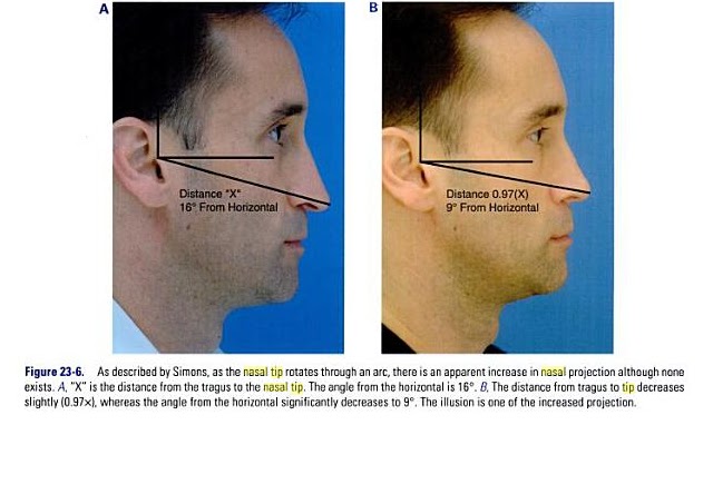
Although nasal tip rotation is not purely a profile adjustment, it is most easily scrutinized from the profile view. Simons describes rotation of the nasal tip as an arc with the radius maintained. [See fig.23-6  Male b/a]  As the tip is rotated, there is some illusion of increased projection although none exists. Tip rotation is defined as the tip angle from the vertical alar crease to the tip. In women, this angle is approximately 105 degrees and in men 100 degrees. The degree of rotation may be affected by the intrinsic properties of the nasal tip (lower lateral cartilages) or external properties (caudal septum).

Nasolabial angle and columella. The nasolabial angle is the angle formed between the columella and upper lip. Ideally, the naslabial angle is 90 to 95 degrees, in men and 95 to 105 in women. [see fig.23-7]

The Nasal Tip as it relates to Profile - Changes in Length, Projection and Rotation

 The position of the nasal tip determines the caudal endpoint of the nose, establishing with the radix nasal length. Changing the projection and rotation of the tip directly affects nasal length. 
 Anderson's tripod principle is most helpful not only in evaluating each tip's unique position but also in guiding what changes will affect tip positon postoperatively. Every step in tip-plasty will somehow affect nasal length, projection, and rotation.  Instead of directly altering the cartilages of the lobule, other more indirect methods to affect relative alar cartilage position include lateral crural overlay or medial crural feet division. These techniques leave the dome area unaffected, whereas affecting the tripod laterally (lateral crural hinge areas) or medially at the feet of the medial crura. A combination of these techniques can create fine changes to tip rotation and nasal length, whereas always decreasing tip projection.

 If the surgical goal is to increase tip projection, then either lateral or medial alar cartilages must be recruited into the lobule area, or onlay cartilage grafts must be added. Lateral or medial crural steal techniques, stabilized by cartilage struts or septal extension grafts, achieve small increases in tip projection. Tip grafts can add substantially more projection, if needed.

Alar Retraction/Hanging
Many expert surgeons now routinely implant small rim grafts at the conclusion of most of their rhinoplasty's to stiffen the alar rim and counteract any tendency for retraction. Occasionally, significant retraction in the revision case will require composite skin-cartilage grafts from the ear to fill the tissue void and scar contracture, especially when it involves the soft tissue triangle. In cases of Hanging ala, rotation changes of the lobule will always improve this subtle deformity.   


Nasolabial Angle

The junction of the columella and lip creates the nasolabial angle; changing its anatomic components can subtlety improve the final rhinoplasty result, uncovering the lip and improving the smile.

Caudal Septum/Spine -{Improve your smile}
The inferior caudal septum and nasal spine comprise the rigid framework for the top of the upper lip. Typically, prominence in this region will increase the nasolabial angle and, of greater esthetic consequence, make the upper lip look pulled up by the nose. Deepening the nasolabial angle by removing inferior cartilage or bone will improve this appearance but may also increase the apparent length of the upper lip. If the nasal depressor muscles are widely detached during this maneuver, the upper lip may also drop, hiding the upper teeth more during smiling. In cases where the smile is already too "gummy" with too much gingival show, this can be a significant  improvement. However, if the upper teeth are already slightly hidden with the preoperative smile, then any muscle detachment should be avoided in this area.

Too acute a nasolabial angle may arise from a deficient premaxilla or too aggressive caudal septal shortening. A premaxillary onlay cartilage graft will help to fill this deficit. Typically, temporary suture fixation of this free graft to the overlying lip helps to keep it in place during the early recovery period.

Columella

 Columellar position is the second component of the nasolabial angle. It may be hanging or hidden.

  Hanging.  A hanging columella is typically seen in two scenarios: either the caudal septum is long, as in a tension nose, or the septocolumellar attachements have been weakened by previous full transfixion rhinoplasty. When the septum is long, shortening it must be accompanied by elevating and reinforcing the medial crura, typically with a strut or tongue-in-groove technique. If the hanging is severe, then a small fusiform excision of membranous columella may also be required. If the septum has been shortened, and the medial crura left unsupported by previous surgery, then re-support with a strut and/or septal extension graft, often with membranous columella excision, will be required.

  Hidden  A hidden columella is typically a postoperative problem, often from over-shortening of the caudal septum or too aggressive a tongue-in-groove technique. A plumping graft that is placed caudal to the medial crura will help to avoid this problem. If unrecognized during an open rhinoplasty, the unsupported open scar will contract during healing, leaving a deficit that will be difficult to correct post-operatively.


It's one year since this blogsite launched

 Time fly's. It's one year since my very first post that launched this site. I actually wasn't planning at that time to do more posts, i simply wanted to make a list of certified Nose Revision Surgeons, explain some do's and don't before agreeing to surgery, and make people aware of risks involved, like neuropathy and empty nose syndrome. I like to take this time to thank you all for coming to my site and for your emails. Each month this site is getting more and more views from Countries all over the world. Majority are from U.S.A. followed by Canada, U.K, Asia, Central Europe. I feel this site is like a grand central station where those contemplating nose revision surgery can feel comfortable to search out the information they're looking for without worries of being solicited; at the same time this site gives exposure as another outlet for Surgeons who write research papers, articles, books and are specializing in Nose Revision Surgery. The more informed you are, the more you'll understand your operated nose, what was performed and why on prior surgery's, and the better your chances at selecting the right Surgeon this time around, of course that's if you decide to go ahead with surgery. Good luck!

Sunday, January 16, 2011

Surgical options for treating "External" Nasal Valve Obstruction

As I mentioned on my last post: Surgical options for treating Internal Nasal Valve Obstruction, there are many surgical options and each surgeon has there favorites. Same applies here for Surgical options for  treating External Nasal Valve Obstruction. The appropriate surgical treatment offered by a surgeon will depend on what is most likely the cause(s), where your obstruction is located and to what degree. Remember to get few opinions on your condition from surgeons that perform a good number of corrective nasal valve surgeries.  I have already posted links on many of the options for correcting external valve collapse on my post titled, Minor Surgical Procedures for treating Nasal Valve Collapse. I will relink them in this post as well.

Rhinoplasty, Postrhinoplasty Nasal Obstruction: Treatment  

Author: Thomas Romo III, MD, FACS Coauthor(s): James M Pearson, MD Paul Presti, MD,Haresh Yalamanchili, MD

External nasal valve obstruction
External nasal valve deformity may be a significant source of nasal airway obstruction in some patients. Constantian and Clardy demonstrated that reconstruction of the external valve alone can improve total mean airflow by more than twice that of preoperative valves.  Interestingly, this degree of airflow improvement is similar to that observed in patients in whom pure internal nasal valve dysfunction was corrected with dorsal or spreader grafts. Moreover, preliminary data for patients in whom both internal and external valve dysfunction were treated (without septal or turbinate surgery) revealed a mean 3-fold airflow increase, which suggests that the effects of internal and external valve reconstruction may be independent but not strictly additive, presumably because of valve interactions.
Static deformity includes tip ptosis and cicatricial stenosis.
  • Tip ptosis, if significant enough, may cause enough narrowing of the vestibule to warrant a tip-lifting maneuver.
    • For structural ptosis, a tip-lifting stitch of 4-0 clear nylon is placed in a horizontal mattress fashion from the dome of the lower lateral cartilage to the periosteum of the nasal bones.
    • For soft tissue ptosis, the thick sebaceous skin at the supratip area along the borders of the nasal subunits can be excised to produce an aesthetically pleasing scar.
    • Tip ptosis is frequently caused by both cartilaginous and soft tissue laxity, as is often observed in the noses of elderly persons (ie, noses affected by aging), and may require a combination of maneuvers.
  • Cicatricial stenosis is another static deformity.
    • Approaches to repair external valve dysfunction secondary to cicatricial stenosis include primary resection, alar interposition, Z-plasty, skin grafts, and composite grafts.
    • Small webs in the external valve area may be divided primarily and then stented. Alar interposition flaps are easily performed but result in increased nasal base width.
Dynamic deformity includes (1) flaccid collapse of lower lateral cartilage after over resection during tip-modeling procedures and (2) nasal musculature deficiency.
  • Flaccid collapse of lower lateral cartilage after over resection during tip-modeling procedures can be corrected with the placement of structural grafts into the alar lobule to provide support and to prevent collapse.
    • Over resection of the caudal upper lateral cartilage and cephalic lower lateral cartilage results in dynamic collapse of the internal nasal valve (upper lateral cartilage) and the external nasal valve (lower lateral cartilage) during inspiration. Dynamic nasal valve collapse (both external and internal) is secondary to a structurally weak or deficient lateral nasal wall.
    • The use of alar batten grafts is an effective method for the correction of internal and external nasal valve collapse secondary to flaccid or absent lateral cartilages. The grafts act to reposition and to provide support to the lateral nasal wall to prevent collapse upon inspiration. They are placed into a precise pocket at the point of maximal lateral wall collapse or supraalar pinching, with the use of either an endonasal or an external rhinoplasty approach.
    • Both septal and conchal cartilages are excellent sources of graft materials. Ensure the graft is of sufficient length to be seated in the soft tissue over the bony piriform aperture. However, it does not need to be particularly long in a caudal/cephalic dimension. After the cartilage is harvested, it is carved into a rectangular shape that spans from the piriform aperture to the junction between the middle and lateral third of the lateral crura. In most cases, these grafts are 10-15 mm long and 4-8 mm wide.
    • To provide maximal structural support, the battens are wider laterally toward the piriform aperture. To minimize cosmetic distortion, battens must be thin with beveled edges, especially along the medial aspect of the graft. Notching the lateral border of the graft helps anchor the batten against the piriform aperture. The size and precise placement of these battens depend on the corrections needed for each individual. Larger grafts are used in patients with severe collapse or thicker skin to provide increased support.
    • The primary purpose of batten grafts is to reinforce areas of the sidewall or the alar lobule that collapse because of the negative force associated with inspiration. Battens are not intended to change the resting position of the valve. Preoperative assessment is critical in the determination of the site of collapse. Once it is identified, a soft tissue pocket is created for placement of the graft. This pocket is usually at the level of the supraalar crease at the junction of the upper lateral cartilage and lower lateral cartilage where previous volume reduction may have been performed. The convex surface of the graft is laterally oriented to provide lateral support for the collapsed region of the lateral nasal wall.
    • In most cases, alar batten grafts create fullness at the site of the graft. This convexity tends to decrease with time as edema resolves and scar contracture compresses the graft and shifts it medially. Andre et al described placement of the graft in a sub-alar position, which has improved cosmetic results but failed to improve symptoms in one-third of patients.
    • The support and stabilization of the lateral nasal wall increases the internal diameter of the nasal airway, thereby increasing dynamic nasal airflow. The increase in size of the internal airway can be appreciated during intranasal examination, with elimination of alar collapse upon moderate-to-deep inspiration. Alar battens are anchored in place with 4-0 chromic gut through-and-through sutures to tack the batten to the nasal mucosa.
    • Positioning of batten grafts may vary from case to case, depending on whether internal or external nasal valve collapse is being treated. When internal nasal valve collapse is treated, battens are typically placed in a pocket at the site of supraalar collapse and are usually near the caudal margin of the upper lateral cartilage or at the point where the lateral crura may have been previously overexerted. When external nasal valve collapse is treated, the grafts are typically placed into a pocket caudal to the cephalically positioned lateral crura.
    • To maximize effect, grafts must be placed into a precise subcutaneous pocket at the point of maximal lateral wall collapse. A tendency is to place the grafts too far cephalically along the upper lateral cartilage, which may result in persistent fullness of the lateral wall of the nose. If the grafts are placed near the alar lobule, they are better camouflaged by the thicker skin. To correct severe nasal valve collapse, the batten grafts can be extended onto the piriform aperture.
    • Toriumi et al reviewed their experience with alar batten grafts in 46 patients and reported that all but one experienced a dramatic improvement in nasal airway obstruction. Postoperative physical examinations revealed a significant increase in the size of the aperture at the internal or external nasal valve. Palpation of the alar sidewalls revealed increased structural support, and examination of the basal view revealed patency of the external nasal valve upon moderate-to-deep inspiration through the nose. Postoperative fullness in the supraalar region in the area where the graft was applied was minimal. With time, this fullness decreased, which left little evidence of the graft and an overall improvement in the aesthetic result.
    • Toriumi et al conclude that alar batten grafts are effective for long-term correction of internal and external nasal valve collapse in patients who do not have intranasal scarring in the region of the nasal valve, loss of vestibular skin, or excessive narrowing at the piriform aperture.
  • With nasal musculature deficiency, severe cases secondary to facial nerve paralysis are treated with dynamic techniques such as nerve grafts, VII-VII crossover, or XII-VII anastomosis.
Rhinoplasty, External Valve Stenosis: Treatment
Author: Alicia R Sanderson, MD Coauthor(s): Craig Cupp, MD, Peter A Weisskopf, MD,

Surgical Therapy

Various surgical options are available to help improve airflow in airways that are obstructed because of a deficient external nasal valve. Options include the following:

  • Septoplasty to address caudal deflection
  • Narrowing a wide columellar base
  • Composite conchal graft to address full- or partial-thickness defects of lateral nasal wall structures secondary to trauma or surgical resection or as a stiffening structure for congenital or senescent weakness1
  • Onlay batten grafts to provide additional lateral wall support
  • Lateral crural strut graft
  • Nasal floor conchal grafts to open an excessively narrowed inferior nares
  • Use of a stitch spreader to help maintain alar stability
  • Reversing the lateral crura
  • Alar expansion stitches with or without alar reinforcement
  • Z-plasty to release internal valve scarring
  • Use of postoperative stents

Preoperative Details

Use the surgeon's standard preoperative preparation for either external or endonasal rhinoplasty. Obtain preoperative photographs. Surgeons should counsel the patient on possible cosmetic changes. Alar batten grafts can result in effacement of the deep alar creases and widening of the nasal tip.

Intraoperative Details

Use the Toriumi method of alar batten graft. The Toriumi method is useful for collapse of external valve secondary to deficiencies of the lower lateral cartilage that lead to collapse. The Toriumi method is not appropriate for absolute narrowing of the piriform aperture or for cases of significant scarring of the valve; however, conchal grafts to the nasal floor or a Z-plasty to release scar may be used in these instances.


Treatment of external nasal valve (alar rim) with an alar strut
A.Kalan, F.R.C.S., G.S.Kenyon, M.D. F.R.C.S. Seemungal, M.R.C.P.
http://ebookbrowse.com/treatment-of-nasal-valve-collapse-with-alar-strut-pdf-d33051866

Surgical management of the septum, turbinates and nasal valve in the treatment of nasal obstruction
Dr. Daniel Becker
http://www.sinustreatmentcenter.com/BOOK/chapter12_0109.pdf

Evaluation of a new procedure for nasal alar rim and valve collapse: Nasal alar rim reconstruction
Robert J. Troell, MD, Nelson B. Powell, MD, Robert W. Riley DDS, MD, and Kasey K.LI, DDS, MD
http://drtroell.com/papers/Nasal_Alar_Rim_Reconstruction.pdf

Lateral Crus Pull-Up
http://archfaci.ama-assn.org/content/8/5/333.full.pdf
Lateral Crural J-Flap
http://oto.sagepub.com/content/128/5/640.full

Narrowing the Columella for (ENVC)
http://spray.me.jhu.edu/~rothbaum/Articles_for_IDP/External%20valve.pdf

Alar Expansion and Reinforcement technique 
http://archfaci.ama-assn.org/content/8/5/293.full.pdf+html 

Plasty techniques
http://www.tantamsj.com/jan2007/30-1-2007.pdf
http://archfaci.ama-assn.org/content/10/3/164.full.pdf
Suspension sutures
http://chicagoent.com/wp-content/uploads/nasalvalvesuspension.pdf

Saturday, January 15, 2011

Surgical options for treating "Internal" Nasal Valve Obstruction

There are many ways to approach surgical correction for nasal obstruction and the aesthetics of the nose. I think of it as a sort of rubiks cube, where you are presented with a unique set of  intermixed colors (representing the patients circumstances they present) which need to be solved by using algorithms and optimally researched proven solutions. Nose Revision Surgeons have their preferences and of course grafts and procedures that are in practice today were developed and popularized by well known Nose Revision Specialist.

Dr. Jack Sheen in 1984 first described spreader grafts as a method of reconstructing the internal nasal valve and/or recontouring the aesthetic appearance of the nasal dorsum in cases of primary and secondary rhinoplasty. Dr. Sheen championed the Closed approach. Dr. Jack Gunter who trained under Dr. Anderson later developed  the lateral crura strut graft and was and still is a devout advocate for the Open approach in secondary rhinoplasty. Dr. Rollin Daniels is credited for developing the technique of diced cartilage wrapped in deep temporal fascia (DC-F). The treatment for Nasal valve collapse largely depends on where exactly the collapse is occurring (can be and usually involves more then one specific area). A competent nose surgeon will know how to properly diagnose the problem and which grafts and/or surgical methods are to be employed to correct not only the breathing issues but also improve the aesthetic results where required. Sometimes more then one procedure may be required, even by different Surgeons, depending on all the issue's presented. The goal is to have a safe long term lasting positive result. Some surgeons prefer to only use your own cartilage, some will recommend irradiated cadaver cartilage, and some like synthetic material or combination of the aforementioned. Furthermore nose revision surgeons who only utilize autologous (from your own body) cartilage grafts have their harvesting site preferences which can be from the septum (if you have enough left and enough can safely remain for the L strut), the ears (auricular grafts), or the ribs (costal cartilage). Some like using bone grafts from different sources and harvest sites for dorsal augmentation. There is no one simple universal answer for all, and you the potential patient will be offered different opinions based on the Nose Revision Specialists training, abilities, experiences and beliefs. This is why it's important for those considering nose surgery to do your due diligence with research. This blogsite should be a big help to most, at least that is my desired intention. I have had 6 surgical nose procedures ( 2 major unsuccessful revisions) and can speak from experience. My main objective for this site is to inform you well enough to make an informed decision so you will avoid the mistakes I have made and therefore hopefully avoid the severe consequences that I ended up with, mostly as a result of my last surgery. I will now focus on some studies that were done on the Surgical Treatments for Internal Nasal Valve Obstruction. If you haven't already read my past posts you may want to see my postings on: Minor Surgical Procedures for treating Nasal Valve Collapse (Jan.10, 2011) , The mystery of all the different types of nose grafts (Oct.18, 2010) and Non-Surgical, Non Medical treatment for collapsed nostrils ( Nasal Valve Collapse ) Oct.31, 2010.
  
Internal nasal valve obstruction 
Rhinoplasty, Postrhinoplasty Nasal Obstruction: Treatment  
Author: Thomas Romo III, MD, FACS Coauthor(s): James M Pearson, MD, Paul Presti, MD, Haresh Yalamanchili, MD 
Static deformity includes: (1) inferomedial displacement of the upper lateral cartilage secondary to hump removal, (2) narrowing of the piriform aperture secondary to osteotomy, (3) scarring at the intercartilaginous junction, (4) turbinate hypertrophy, and (5) deviated nasal septum.
  • Inferomedial displacement of the upper lateral cartilage secondary to hump removal can be treated with spreader grafts, flaring sutures, butterfly grafts, or a combination thereof.
    • Methods of correcting internal nasal valve collapse are focused on the reposition of the upper lateral cartilage or the addition of structural grafts to support the lateral wall of the nose. Spreader grafts are often used for the correction of internal nasal valve collapse. These grafts reposition the upper lateral cartilage in a lateralized position and add width to the middle nasal vault. Numerous other structural grafts have been described as providing support to the lateral nasal wall. Most support the weakened upper lateral cartilage and lateralize the caudal margin of the upper lateral cartilage at the nasal valve. The techniques of repair differ among different authors and can be achieved via either the open or endonasal approach.
    • Spreader graft placement is the workhorse repair of the narrowed internal nasal valve.
      • These grafts are designed to lateralize the upper lateral cartilage by the width of the graft, thereby increasing the cross-sectional area of the nasal valve.
      • Septal cartilage can be harvested and shaped into spreader grafts. If the septum is unavailable, conchal cartilage or Medpor may be used. {For some reason the authors don't mention costal cartilage (rib) grafts which in the hands of a competent surgeon is a excellent choice}. I personally do not like the idea of Medpor which is a synthetic material.
      • The grafts are placed in a submucosal pocket between the septum and the upper lateral cartilage. These grafts are typically 1-2 mm thick and extend the entire length of the upper lateral cartilage from the cephalic border beneath the nasal bones to the caudal margin. They are anchored in place with one or two 5-0 polydioxanone horizontal mattress sutures that span from one upper lateral cartilage through the ipsilateral spreader graft, the septum, the contralateral spreader graft, the contralateral upper lateral cartilage, and then back again.
      • In a series of 29 patients with pure internal valvular incompetence treated with spreader grafts alone, Constantian and Clardy reported a 2-fold increase in postoperative airflow.
      • Andre et al reported significant improvement in nasal airway patency with autologous endonasal spreader grafts. A total of 89 patients, at an average follow-up of 12.2 months, were reviewed for symptomatic improvement of their nasal obstruction after placement of spreader grafts. Most (88%) had favorable results. Their technique involved placement of the graft within a tight-fitting subperichondrial pocket between the nasal septum and the upper lateral cartilages. Fixation of the grafts was provided via suturing, tissue glue, or simply the tension of a tight pocket.
    • Flaring sutures are a simple way to improve the cross-sectional area of the internal nasal valve by directly changing the internal valve angle.
      • Although the spreader graft moves the dorsal border of the upper lateral cartilage in a lateral direction, the angle of the internal valve is minimally affected.
      • A 4-0 polydioxanone horizontal mattress stitch extends from the caudal/lateral area of the upper lateral cartilage and across the dorsum of the nose and is anchored to the contralateral upper lateral cartilage. As the suture is tightened, both upper lateral cartilages are pulled laterally, with the dorsum serving as a fulcrum. This flaring action directly affects the internal valve angle, and its effects can be witnessed as the suture is tightened.
      • When used in conjunction with spreader grafts, the focal point of the flaring suture is moved laterally to a more optimal position. The addition of a flaring suture to conventional spreader graft placement is simple and quick and dependably improves treatment of the dysfunctional internal nasal valve. Both flaring sutures and spreader grafts serve to move the upper lateral cartilage to a lateral and externally rotated position.
    • An alternative to the flaring suture is the placement of a 3-0 Prolene suspension suture.
      • Rizvi and Gauthier published their experience with this technique in 40 patients with internal nasal valve collapse. Over a follow-up period of 2-3 years, all of the patients reported improvement of their nasal obstruction.
    • Butterfly grafts take advantage of the intrinsic curvature of conchal cartilage to improve the nasal airway.
      • The grafts may be placed endonasally or with an open approach. They are placed at the scroll area between the upper lateral cartilage and lower lateral cartilage in an attempt to widen the valve angle. The caudal border of the graft may be placed deep to the cephalic border of the lateral crura to help camouflage the graft.
      • Grafts are anchored in place with 5-0 polydioxanone to prevent migration.
      • Butterfly grafts, more than other valve-plasty maneuvers, can lead to postoperative cosmetic changes, with marked fullness along the supratip area.
  • Narrowing of the piriform aperture secondary to osteotomy can be treated with revision osteotomy with outfracture of the nasal bones to widen the valve angle.
    • Occasionally, severe valve narrowing occurs after rhinoplasty as a result of lateral osteotomy with infracture, which may not improve with any of the aforementioned procedures. These patients can be treated with revision osteotomy with outfracture of the nasal bones to widen the valve angle. The revision lateral osteotomy is made in the same line as the original osteotomy in order to mobilize the bone that was displaced too far medially. The frontal process of the maxilla and the nasal bones are then lateralized (outfractured).
    • Outfracture can adversely affect cosmesis by widening the nasal dorsum. Therefore, attempt more conservative approaches initially; however, revision osteotomy with outfracture may still be an option to correct significant nasal breathing dysfunction due to valve narrowing after rhinoplasty.
    • Pontell et al compared the cross-sectional area between infracture with the outfracture position and noted an increase of more than 200% with outfracture.14 A change of 1° in valve angle increased the area by approximately 4 mm2.
  • Scarring at the intercartilaginous junction can be treated with scar excision.
    • Scarring at the valve apex or valve angle can be corrected with scar excision followed by reconstruction with the use of a full-thickness skin graft or local mucosal flap. Finding enough adjacent unscarred lining, skin, or mucous membrane to effectively correct a contracture blunting the apex of the valve is usually difficult.
    • Full-thickness skin grafts and composite grafts are reasonable for the reconstruction of small defects. Full-thickness skin grafts are taken from the upper eyelid or postauricular area. Carefully fit these grafts into the defect using 5-0 or 6-0 absorbable sutures.
    • Larger scars or webs require Z-plasty, V- to Y-plasty, or mucosal advancement flaps from the septum or labial mucosa.
  • Deviated nasal septum can be treated with septoplasty.
    • Septal abnormalities probably represent the most frequent cause of nasal valve obstruction. The septal cartilage, bone, or both may be thickened, be deflected off the nasal spine, be twisted, be scarred, have spurs, or be affected by a combination of these.
    • A septal abnormality that occurs at the nasal valve area can produce nasal airflow obstruction, which can be corrected with the performance of a septoplasty.
Dynamic deformity includes dynamic collapse of the upper lateral cartilage.The dynamic collapse of the internal nasal valve during inspiration secondary to an unsupported upper lateral cartilage can be corrected with the placement of a butterfly graft or a batten graft at the scroll area (caudal aspect of the upper lateral cartilage) to give support to this critical area. Alar batten grafts can be used to correct internal or external nasal valve collapse.
  • For internal nasal valve collapse, place the alar batten grafts in a precise pocket at the point of maximal lateral wall collapse. This point is usually near the caudal margin of the upper lateral cartilage and cephalic margin of the lateral crura of the lower lateral cartilage, where previous volume reduction may have been performed.
  • Spreader grafts can be used in combination with alar batten grafts when excessive narrowing of the middle nasal vault is present.
  • The use of alar batten grafts is discussed in further detail in the section regarding the correction of dynamic external nasal valve collapse after rhinoplasty secondary due to overresection of lower lateral cartilage
http://emedicine.medscape.com/article/841574-treatment
Rhinoplasty, Internal Valve Stenosis: Treatment   
Author: David Núñez-Fernández, MD, PhD Coauthor(s): Jan Vokurka, MD, PhD,Gloria Fernández-Muñoz, MD

Surgical Therapy

Several techniques are used to correct a stenotic or collapsed nasal valve. Depending on the type of pathology, the surgeon can choose to use one or several methods. The scope of techniques varies from sutures to the application of grafts. The common goal is to open the valve, restoring the appropriate anatomy. Explaining each technique is beyond the scope of this article; however the following is a summary of these techniques depending on the structure modified.

A valvuloplasty is the surgery historically used to reconstruct the nasal valve. The goal of this surgery is to open the valve by removing the returning ULC and trimming the caudal border of the cartilage. It is not the only technique used to correct valvular alterations, but it provides an excellent view of the caudal border of the ULC and can be used in conjunction with other techniques. Because the valve is formed by several different structures, other techniques may be necessary, depending on the type of deformity that produced the stenosis.

Conchal cartilage butterfly graft
This graft has been found useful by Friedman and Cook in primary rhinoplasty. It has been used traditionally for secondary surgery when too much ULC has been resected. The natural convexity and rigidity of the conchal cartilage is an excellent option to open the ULC.

Intraoperative Details
Because the valve is formed by several structures, the surgery is directed toward realigning the obstructing parts. The authors discuss treatment of the Septum, Nasal roof, Upper and Lower cartilage, Inferior Turbinates, Pyriform Aperture, Synechia or scarring of the mucosa of the valve.

Note: I personally do not agree with their recommendation of removing the anterior "head" portion of the inferior turbinates up to 2cm. There are dangers of ending up with Empty Nose Syndrome especially if you previously had a turbinate procedure.  So keep in mind some of the mentioned  options are safer then others. 

http://emedicine.medscape.com/article/877468-treatment

Turbinate Surgery
 The turbinates provide much of the good, disease-fighting stuff in the nasal cavity and sinuses: mucus, cilia, and enzymes (such as lysozyme). If the inferior and middle turbinates are removed, the source of warming and moistening the air is gone, and then the dryness, frequent infections, crusting and local pain can result. With the absence of the mucus, cilia and enzymes, frequent infections of the sinuses can occur. This may lead to the need for further sinus surgery. 

Two methods that are safe and don't cause major harm to the cila and mucosa of the turbinates in your nose, are microdebreider assisted turbinate surgery, and somnoplasty.  They are safer then cauterizing the turbinates or using laser heated techniques.  Cutting out the turbinates could lead to serious consequences such as empty nose syndrome.  Sometimes it may be necessary to remove small portion of the turbinate bone- submocosus turbinate reduction, but preserving the mucosa is still critical for a healthy nose. 

Procedures that may result in Empty Nose Syndrome when done improperly include wide chemical or electric cautery, laser cautery, and of course, surgical removal. 
Dr. Murray Grossan http://www.ent-consult.com/emptynose.html#surgeries 
Dr. Steven Houser    http://webspace.webring.com/people/jn/nares/turb.html  
Sinus, Polyps and other types of obstructions http://www.obstructednose.com/index.html 

Nasal Valve Reconstruction

[Experience in 53 Consecutive Patient]

Authors:Maurice M. Khosh, MD Albert Jen, MD; Carlo Honrado, MD;Steven J. Pearlman, MD

Results  The most common cause of nasal valve obstruction was previous rhinoplasty (79%), followed by nasal trauma (15%) and congenital anomaly (6%). Spreader grafts were used in 42 patients (79%), and alar batten grafts were used in 19 patients (36%). The patients received a minimum of 1 year of follow-up. All 12 patients with external valve dysfunction showed improvement after surgery. Thirteen (93%) of the 14 patients with concomitant external and internal valve dysfunction had improvement in nasal obstruction after treatment. Twenty-four (89%) of 27 patients with internal nasal valve dysfunction reported improvement in nasal obstruction. Spreader grafts caused a widening of the middle third of the nose. Alar batten grafts resulted in effacement of deep alar creases and a widening of the nasal tip.
Conclusions  We have found that surgical correction of nasal valve obstruction is extremely effective in improving subjective nasal obstruction. Success of this procedure is predicated by correct diagnosis and appropriate surgical technique. 


For additional information see the following links:
http://www.naturalrhinoplasty.com/html/functional_rhinoplasty.php
http://www.ncbi.nlm.nih.gov/pubmed/16732504
http://emedicine.medscape.com/article/1292527-overview
http://findarticles.com/p/articles/mi_m0BUM/is_3_83/ai_n6077596/

Wednesday, January 12, 2011

Septal cartilage (quadrangular cartilage) and the L strut

The cartilage of the septum (or septal cartilage, or quadrangular cartilage) is somewhat quadrilateral in form, thicker at its margins than at its center, and completes the separation between the nasal cavities in front.
Its anterior margin, thickest above, is connected with the nasal bones, and is continuous with the anterior margins of the lateral cartilages; below, it is connected to the medial crura of the greater alar cartilages by fibrous tissue. Its posterior margin is connected with the perpendicular plate of the ethmoid; its inferior margin with the vomer and the palatine processes of the maxillae.

In regards to grafting, autogenous septal cartilage is considered the Gold Standard

The term L strut can refer to the specific area in the quadrangular cartilage region that remains  after septal cartilage is removed from the area normally for grafting purposes. It can also refer to a synthetic L shaped implant. Sometimes if too much cartilage is removed from the septal quadrangular region then reconstruction is needed to maintain the sacrosanct L strut for fundamental support of the nose.  

 Dr. Naderi mentions in his blog: Septal cartilage: A large portion of the Septal Quadrangular Cartilage can be harvested and used during Rhinoplasty. “An L-strut” must be left to support the nose. This L-strut must be at least 1.5cm in width to support the bridge and tip. ( Note: That is general standard accepted in the practice )The remainder of the cartilage can be removed and used for Rhinoplasty. In Revision Rhinoplasty, this cartilage is often missing and unavailable, as it may have been used during the previous Rhinoplasty surgeries. Septal perforation (hole) is a risk of septal cartilage harvest or septoplasty. This cartilage is often missing and destroyed in patients who have been struck to the nose very hard (boxers) as well as patients with history of Cocaine abuse or autoimmune diseases.

Role of the Nasal Septum
From Head and Neck surgery by Dr. Byron J. Bailey and Jonas. T. Johnson

The combination of the cantilevering dorsal element and the buttressing caudal element forms the basis of the L-shaped strut- the most structurally important aspect of the quadrangular cartilage. Compromise to the dorsal component leads to a saddle nose deformity with ventral collapse of the upper cartilaginous vault. The classic example of this is quadrangular cartilage resorption after an untreated septal hematoma. Compromise to the caudal component may lead to nasal tip ptosis, particularly in the presence of weak medial crura. Tramatic or iatrogenic injury is most often the cause. For these reasons, it is critical to maintain structural integrity of the L-strut during surgery. Although conventional teaching emphasizes the need to maintain an uninterrupted 1.5 cm wide dorsal-caudal strut, malposition or deformity of the strut itself may be the cause of external deformities, particularly in the crooked nose. Correction of these problems may mandate the use of techniques that modify the septum and may require repositioning, camouflaging, or reconstruction of the L-struct itself.

Now, here's information on Synthetic L implant.
 Nasal Implants: Is an I-shaped implant better than an L-shaped implant?
 By Drs.Litner and Solieman

How about neither? We are often asked about nasal implants. These are primarily used for East Asians, African Americans, and others seeking a stronger, higher nasal bridge and a more refined tip. They are also sometimes recommended for patients whose bridge has been lowered too much during previous surgery. Implants can be of various materials but the most commonly used implants are made of silicone/silastic.


nasal implant
Above you can see the typical shape of a nasal L-strut implant. This particular one happens to made of Medpor which can be a real problem to revise

An L-strut lays over the entire bridge and extends down under the tip to the base of the columella. It gives the tip definition by placing a fair bit of pressure over a small area of skin to tent the skin out. It cannot reproduce or retain the natural shape of your tip. As a result, it can look a little too pointy and unnatural.Sometimes, this pressure on the skin can become too much for the skin to bear.

Our preference for ethnic augmentation rhinoplasty is to use your own cartilage for augmentation.

Monday, January 10, 2011

Minor Surgical Procedures for treating nasal valve collapse (NVC)

  A  variety of literature on minor surgical  procedures happens to exist claiming high efficacy for those suffering with nasal valve collapse. But chances are you  never heard about most of them. Some of the specific procedures are used in conjunction with other surgical grafting procedures, but a number of studies have shown that these procedures exclusive to other procedures can stand on their own for remedying NVC  for certain individuals that are diagnosed correctly.  They don't involve major grafting of the nose, or any in most cases, relatively not intricate and can take anywhere from 1-2 hours to complete,  could be covered by insurance or Government health care system, can improve nose aesthetically or remain about the same,  and very minimal down time. Caution:  Nasal valve collapse is a multidimensional problem. In some patients, the reduced cross-sectional area or an acute valve angle of less than 10° is the main problem. In others, the weak nasal sidewall plays a major role. In some patients, a combination of factors exists.Consequently, because most of these procedures focuses on only 1 factor, none is universal for every situation.

I will divide some of these procedures as follows: Plasty techniques, Suture techniques, Lateral crura options, butterfly grafts and synthetic injectable  grafts. I plan to discuss most if not all in more detail in upcoming posts.

Here's a list of  minor procedures for improving NVC.

Suture Techniques
  • Flaring Sutures
  • Nasal Valve Suspension
http://chicagoent.com/wp-content/uploads/nasalvalvesuspension.pdf

 Plasty Techniques 
  • M-Plasty technique
  • Z Plasty technique
http://www.tantamsj.com/jan2007/30-1-2007.pdf
http://archfaci.ama-assn.org/content/10/3/164.full.pdf
    Butterfly grafts
    • autologous grafts from ear
    • Titanium expanded polytetrafluoroethlene e-PTFE
    http://www.nc-faces.com/the-butterfly-graft/
    http://archfaci.ama-assn.org/content/8/2/98.full.pdf

    Alar Expansion and Reinforcement technique 
    http://archfaci.ama-assn.org/content/8/5/293.full.pdf+html 

    Lateral crura options    (Some of these graft procedures are performed together or in combination with other procedures and therefore considered Major Nose Revision Surgery)
    •  Lateral strut grafts
    •  Flip Flop grafts
    • Alar batten grafts
    • Alar rim grafts
    • Lateral Crus Pull-up 
    • Lateral Crural J-Flap
    http://drtroell.com/papers/Nasal_Alar_Rim_Reconstruction.pdf
    http://archfaci.ama-assn.org/content/8/5/333.full.pdf
    http://oto.sagepub.com/content/128/5/640.full
      Revision Osteotomy with outfracture of nasal bone

      Narrowing the Columella for (ENVC)
      http://spray.me.jhu.edu/~rothbaum/Articles_for_IDP/External%20valve.pdf

      Injectable spreader grafts  *note
      • hydroxyapatite (Radiesse)
      • hyaluronic acid (Restylane)
      http://findarticles.com/p/articles/mi_m0BUM/is_5_86/ai_n27270780/
      *note
      Dr. Dean Toriumi has reported serious issues with injectable fillers in the nose. Here is some information from his website on this issue.
      The most concerning phenomenon is that many dermatologists, generalists and surgeons are performing non-surgical rhinoplasties. In these cases, semi-permanent or permanent injectable filler materials are being injected into the nose to make long lasting contour changes. Some of those who are performing the injections are not rhinoplasty surgeons and may have little if any understanding of the nasal anatomy and nasal aesthetics. I have seen many patients treated by such physicians with severe nasal skin envelope problems such as infection, swelling, pain, permanent redness and deformity. Unfortunately, many of these patients cannot be helped because correction of the problem requires resection of the filler material. Resection puts these patients at severe risk of permanent skin damage in the form of intense redness or skin necrosis, leaving a hole in their nose. Additionally, we do not know the long term effects of such materials on the nasal skin envelope.

      There is an intense need for scientifically sound research that demonstrates the safety and efficacy of these materials in the nose. Scientific research may show that these filler materials when placed deeply against the bone and cartilage are safe when used in the nose. The nose is a very important structure of the face that greatly influences the overall facial appearance. Caution should be taken when doing anything that could potentially damage the nose and leave the patient with a permanent deformity.

      Confused about what's the difference between Lateral crural strut grafts and alar strut grafts? Or rim grafts and Alar batten grafts?
      Dr. Naderi explains in simple terms. Alar strut grafts and Lateral crural strut grafts are same.  But Alar batten grafts are not the same. A "rim graft" sits along the alar rim (right at the margin just like it sounds) whereas a "alar strut graft" site higher OVER the lateral aspect of the Lower Lateral Cartilage. The rim graft supports the soft tissue rim (nostril) while the strut graft supports the LLC laterally and thereby supports the entire nostril sidewall, not just the rim. An alar batten graft is a longer grafts that sits UNDER the lateral aspect of the Lower Lateral Cartilage and extends to the body pyriform apperture to prevent collapse of the nostrils during forceful inspiration.