Showing posts with label nasal tip. Show all posts

Saturday, December 10, 2011

Refining the nasal tip with Newer VDD techniques.

Vertical Dome Divison Rhinoplasty: 
 Author: John Hilinski, CoAuthors: Anil R. Shah & Kris S Moe

Nasal tip surgery is among the most complex and difficult tasks in rhinoplasty surgery. Vertical dome division (VDD) is one of a variety of techniques that may be used in refining nasal tip appearance. Collectively, vertical dome division (VDD) refers to one of many methods of vertically dividing the lower alar cartilage at or near the dome to modify nasal tip aesthetics. The technique was originally recommended as an alternative in altering tip projection and appearance while minimizing use of implants and the degree of postoperative tip ptosis. Vertical dome division (VDD) targets various nasal deformities, including overprojection or underprojection, suboptimal rotation, disproportionate lobule ratios, and broad or asymmetric tip. The surgeon must strive to achieve an aesthetically pleasing nasal tip that is in balance with the remainder of the nose without compromising nasal airway function.

The typical patient presenting for vertical dome division (VDD) usually has a poorly defined or malpositioned tip with a combination of abnormal projection/rotation, broad or amorphous lobule, asymmetric tip defining points, and/or boxy, trapezoidal base.

Adherents to this principle argue that horizontal excisional techniques rely too heavily on unpredictable and uncontrollable postoperative scarring to produce desired tip results. Proponents of vertical dome division (VDD) believe that vertical incisional and excisional techniques, on the other hand, offer a more definitive and reliable means to achieve desired tip changes.

In principle, the technique of vertical dome division (VDD) separates the medial and lateral crura into 2 independent units. By transecting the dome, the inherent spring within the arch is released and allows realignment of the newly divided medial and lateral segments to reconstruct the nasal tip.

Vertical dome division (VDD) is typically reserved for more complicated cases that require greater changes to effect tip refinement than could be achieved using other techniques. Nearly all variations of vertical dome division (VDD) used today involve some modification of the original Goldman technique

The lobule is defined as the portion of the nasal tip complex that is situated anterior to the nostrils; it extends from the tip defining point to the junction with the columella, as observed on base view. The alar cartilage (lower lateral cartilage) is C-shaped and can be divided into the medial, middle, and lateral crus. The middle (intermediate) crus comprises the domal segment and largely influences the shape of the lobule and, therefore, the form and definition of the nasal tip. The dome is considered the highest arching segment within the nasal vestibule.

The lobule size can be assessed in comparison to the columellar length. If the base view demonstrates a columellar-to-lobule ratio of approximately 2:1, the structural support and configuration of the nasal tip is considered adequate. A long nasal length reflects an acute nasolabial angle; a short length reflects an obtuse nasolabial angle.

The anatomy of the nasal tip is often described using the tripod concept to facilitate understanding of the key structural components and to provide a simple explanation of tip dynamics. According to this analogy, the cartilaginous framework of the lower third of the nose is compared to a tripod that is attached to the facial frontal plane. The 2 individual lateral crura represent 2 legs of the tripod, and the conjoined medial crura and caudal septal attachments correspond to the third leg.

By lengthening or shortening any or all legs of the tripod, the changes that will be effected in tip projection and rotation can be predicted. For instance, techniques that augment or lengthen the medial crural segment enhance projection. Shortening the medial crura or disrupting their septal attachments without reduction of lateral crural length decreases projection and rotation of the nasal tip. Shortening the lateral crura and maintaining or lengthening the medial crural segment would be expected to increase rotation.

Contraindications

Vertical dome division (VDD) is predominantly contraindicated in patients with relatively thin skin. These patients are particularly prone to developing visible cartilage edges along the nasal tip region. This results from contraction of the thin overlying skin and soft tissue envelope around the new and more prominent medial cartilaginous strut. A thick overlying skin and soft tissue envelope is better able to cushion the appearance of prominent cartilaginous structures, such as those in vertical dome division (VDD).

Avoid classic vertical dome division (VDD) in patients who show evidence of already weakened lateral nasal walls. Dividing the domal region without reapproximation of the cartilage segments disrupts the integrity and continuity of the lower lateral cartilage. The lateral nasal wall is more susceptible to structural collapse than the newly reinforced medial footplates. As a result, lateral wall weakening and collapse are further potentiated.

The original technique is highly focused on manipulation and repositioning of only the medial crura, with no attempt made to reconstruct the remaining lateral crural segment and alar rim. Postoperatively, the medial crura and columella are sufficiently stable to resist loss of projection. Some loss of lateral support, which could result in lateral wall collapse and alar retraction, may occur. Vertical dome division (VDD) is also associated with bossae formation. This is most frequently seen in patients with thin skin and firm cartilages. Disruption of the underlying vestibular mucosa and skin, such as in the Goldman technique, also predisposes the patient to possible stenosis.

With use of newer modified techniques (Hockey stick excision, Lipsett, Simons, Adamson, and others) and caution, vertical dome division (VDD) can be used successfully in nasal tip refinement with limited postoperative complications and reliable long-term results. 

Most practitioners using vertical dome division (VDD) today perform some modification of the original Goldman tip procedure.

Adamson reported that approximately 5% of patients required revision surgery for postoperative tip abnormalities and irregularities attributed to use of vertical dome division (VDD).[10] These abnormalities were primarily nasal bossae and lobule asymmetries. Abnormalities were nearly 3 times as likely to occur in revision cases as in primary rhinoplasty; incidence was lower with use of the incision and overlap method.

 Vertical dome division (VDD) is likely to remain controversial in the future. As knowledge of nasal tip surgery evolves, so too will further modifications of our existing tip techniques to attain more predictable outcomes.
 http://emedicine.medscape.com/article/841313-overview

The Endonasal Approach to Rhinoplasty
Robert L. Simons and Lisa D. Grunebaum Ch.68 Rhinology and Facial Plastic Surgery 

Today's trends in nasal tip surgeries are for improved visualization with preservation of the cartilage as well as better medial stabilization and support. These tenets are inherent in vertical dome division techniques. It is important to remember that VDD is an incisional technique that allows for repositioning of the nasal tissue and should not be combined with excisional techniques. One should always leave behind more than one takes. Preservation of at least 6-8mm of lateral crus will help prevent alar collapse and help stabilize the nasal base as well as allow for a strong natural-appearing tip.VDD allows for narrowing, rotation, and change in tip projection by repositioning rather than excision of any sizable amount of cartilage.


The Effectiveness of modified VDD Technique in reducing nasal tip projection in rhinoplasty
B.Gandomi, M.H. Arzaghi, M. Rafatbakhsh

The employed technique is a new modification of previous techniques. 3-10 The technique employs an open approach in which a strong columellar strut inserted and a portion of crura is removed near the dome (the cornerstone of our new technique of tip surgery). Depending on the deformity, this segment may involve intermediate, middle or lateral crura with or without removal of vestibular skin considering its thickness: thin skins are not resected, but thick skins are usually removed.

Our technique involves the overlapping of the incised edges of the medial and lateral segments, and suture approximation to restore the integrity of the alar cartilage. The technique allows a more stable configuration for the maintenance of nasal tip support. The overlapping and reapproximation of the medial and lateral units ensure the long-term stability of the newly reconstructed nasal tip complex, and reduce the tendency towards postoperative cartilaginous abnormalities that may accompany scar fibrosis and contracture. 


Wednesday, January 19, 2011

The Nose Tip: Dimensional Analysis 101

Advanced Therapy in Facial Plastics and Reconstructive Surgery  By: Dr. Regan Thomas

Tip Support Mechanisms
 Nasal tip shape is largely determined by the shape and integrity of the LLC (lower lateral cartilage). The tip surface is divided into the dome, alae, soft triangles, and columella. The major tip support mechanisms include the size, shape and integrity of the LLC, the attachment of the medial crus to the septal cartilage, and the attachment of the LLC to the ULC (upper lateral cartilage).  The minor support mechanisms are the interdomal ligament, dorsal portion of anterior septal angle, sesamoid cartilages, the LLC attachment to skin/soft tissue, and the nasal spine.


 http://www.lamfacialplastics.com/resources/learning-modules/rhinoplasty-tutorial/

Nasal Profile Projection   Minas Constantinides M.D. and Michael Carron M.D.
Unlike nasal length, which is relatively difficult to change, nasal projection can be changed easily. Because of this ease, however, changes are often overdone. Secondary corrections of overdone projection changes are among the biggest challenges faced in revision rhinoplasty.  Projection of the tip is the actual measured distance from the alar-facial plane to the tip. A change in nasal projection requires either an increase or decrease in the distance the nasal tip extends from the vertical facial plane. The Goode method and the 3:4:5 triangle are the two most common ways of measuring projection. [See Fig.23-4, Fig 23-5]

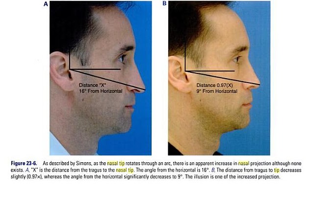
Although nasal tip rotation is not purely a profile adjustment, it is most easily scrutinized from the profile view. Simons describes rotation of the nasal tip as an arc with the radius maintained. [See fig.23-6  Male b/a]  As the tip is rotated, there is some illusion of increased projection although none exists. Tip rotation is defined as the tip angle from the vertical alar crease to the tip. In women, this angle is approximately 105 degrees and in men 100 degrees. The degree of rotation may be affected by the intrinsic properties of the nasal tip (lower lateral cartilages) or external properties (caudal septum).

Nasolabial angle and columella. The nasolabial angle is the angle formed between the columella and upper lip. Ideally, the naslabial angle is 90 to 95 degrees, in men and 95 to 105 in women. [see fig.23-7]

The Nasal Tip as it relates to Profile - Changes in Length, Projection and Rotation

 The position of the nasal tip determines the caudal endpoint of the nose, establishing with the radix nasal length. Changing the projection and rotation of the tip directly affects nasal length. 
 Anderson's tripod principle is most helpful not only in evaluating each tip's unique position but also in guiding what changes will affect tip positon postoperatively. Every step in tip-plasty will somehow affect nasal length, projection, and rotation.  Instead of directly altering the cartilages of the lobule, other more indirect methods to affect relative alar cartilage position include lateral crural overlay or medial crural feet division. These techniques leave the dome area unaffected, whereas affecting the tripod laterally (lateral crural hinge areas) or medially at the feet of the medial crura. A combination of these techniques can create fine changes to tip rotation and nasal length, whereas always decreasing tip projection.

 If the surgical goal is to increase tip projection, then either lateral or medial alar cartilages must be recruited into the lobule area, or onlay cartilage grafts must be added. Lateral or medial crural steal techniques, stabilized by cartilage struts or septal extension grafts, achieve small increases in tip projection. Tip grafts can add substantially more projection, if needed.

Alar Retraction/Hanging
Many expert surgeons now routinely implant small rim grafts at the conclusion of most of their rhinoplasty's to stiffen the alar rim and counteract any tendency for retraction. Occasionally, significant retraction in the revision case will require composite skin-cartilage grafts from the ear to fill the tissue void and scar contracture, especially when it involves the soft tissue triangle. In cases of Hanging ala, rotation changes of the lobule will always improve this subtle deformity.   


Nasolabial Angle

The junction of the columella and lip creates the nasolabial angle; changing its anatomic components can subtlety improve the final rhinoplasty result, uncovering the lip and improving the smile.

Caudal Septum/Spine -{Improve your smile}
The inferior caudal septum and nasal spine comprise the rigid framework for the top of the upper lip. Typically, prominence in this region will increase the nasolabial angle and, of greater esthetic consequence, make the upper lip look pulled up by the nose. Deepening the nasolabial angle by removing inferior cartilage or bone will improve this appearance but may also increase the apparent length of the upper lip. If the nasal depressor muscles are widely detached during this maneuver, the upper lip may also drop, hiding the upper teeth more during smiling. In cases where the smile is already too "gummy" with too much gingival show, this can be a significant  improvement. However, if the upper teeth are already slightly hidden with the preoperative smile, then any muscle detachment should be avoided in this area.

Too acute a nasolabial angle may arise from a deficient premaxilla or too aggressive caudal septal shortening. A premaxillary onlay cartilage graft will help to fill this deficit. Typically, temporary suture fixation of this free graft to the overlying lip helps to keep it in place during the early recovery period.

Columella

 Columellar position is the second component of the nasolabial angle. It may be hanging or hidden.

  Hanging.  A hanging columella is typically seen in two scenarios: either the caudal septum is long, as in a tension nose, or the septocolumellar attachements have been weakened by previous full transfixion rhinoplasty. When the septum is long, shortening it must be accompanied by elevating and reinforcing the medial crura, typically with a strut or tongue-in-groove technique. If the hanging is severe, then a small fusiform excision of membranous columella may also be required. If the septum has been shortened, and the medial crura left unsupported by previous surgery, then re-support with a strut and/or septal extension graft, often with membranous columella excision, will be required.

  Hidden  A hidden columella is typically a postoperative problem, often from over-shortening of the caudal septum or too aggressive a tongue-in-groove technique. A plumping graft that is placed caudal to the medial crura will help to avoid this problem. If unrecognized during an open rhinoplasty, the unsupported open scar will contract during healing, leaving a deficit that will be difficult to correct post-operatively.


Thursday, January 6, 2011

Introduction to M-Arch model and Tripod theory application for the nasal tip

The M-Arch Model: A New Concept of Nasal Tip Dynamics
Adamson, P.A., Litner, J.A., Dahiya, R. | Arch Facial Plast Surg | vol. 8, 16 - 25, 2006 

Background: 
Of the many techniques for addressing the nasal tip, the tripod concept of nasal tip dynamics is one that has stood the test of time. This concept defines the conjoined medial crura as 1 leg of a tripod and each lateral crura as the other legs. The lengths of these legs can be adjusted to alter the tip position. The M-Arch model extends this concept to think of the tripod as an arch with a specific length that can be manipulated anywhere within the arch. Thus, length, projection, rotation, and lobule refinement can be achieved through technical maneuvers performed in a graduated and integrated approach. The M-Arch model also defines the domal arch, a lobular-arch consisting of the intermediate crura and anterior component of the lower lateral crus. A key component in making changes in length, projection, rotation, and lobule refinement is vertical division, specifically, making an incision in the tripod arch that is perpendicular to the long axis of the arch. Vertical divisions can also be used to alter columellar, lobular, and lower lateral crural deformities and change the position of the tip-defining point.

Applications: 
The M-Arch model can be used to change tip projection. Increased tip projection is achieved by increasing tripod arch length anteriorly. Decreased tip projection is achieved by performing a vertical arch division and/or excision at the medial and lateral crural feet. The relative effect on projection and rotation is determined by the placement of the vertical lobule division. Counter-rotation of the tip-defining point occurs when the medial crura are shortened more than the lateral crura. If the reverse is performed, rotation is produced. Rotation shortens the nose; counterrotation lengthens it. With respect to lobule definition, vertical lobular division in the intermediate crus can be a significant tool to address deprojection, rotation, and lobular refinement. It is also useful in diminishing a hanging infratip lobule, narrowing a broad biconvex domal arch, and improving nostril-lobular relationship when the infratip lobule height is relatively large. Various other uses are possible. Ultimately, using the M-Arch model helps the surgeon produce the desired esthetically pleasing nasal tip.


Rhinoplasty, Tripod Theory

Author: Anil P Punjabi, DDS, MD

Introduction

Surgically manipulating the nasal tip to achieve predictable results is the most difficult feature of rhinoplasty. One who can control the nasal tip is said to be able to master rhinoplasty. Anderson first proposed the rhinoplasty tripod theory. A complete comprehension of the tripod theory and the dynamics of tip projection, support, and rotation allow categorization of the different factors that may need alteration to manipulate the nasal tip.
Rhinoplasty is technically demanding, and tip surgery is an art form. It requires an in-depth knowledge of the complex three-dimensional anatomy of the nose, a complete understanding of its physiology, familiarity with the described techniques of nasal tip surgery, and a well-developed sense of aesthetics; all are essential in mastering rhinoplasty.

The Tripod Theory
The tripod concept of tip projection, support, and rotation described by Anderson provides an understanding of the dynamics of tip rhinoplasty. The anatomy of the two alar cartilages forms a functional tripod that provides tip support. The right and left lateral crura comprise two legs of the tripod, and two conjoined medial crura function as the third leg. Anatomically, the medial crura are shorter than the lateral crura. The medial crural foundation is supported by the attachments to the superior and inferior septum.

Incisions between the upper and lower lateral cartilages (ie, intercartilaginous incisions) and removal of the cephalic border of the lateral crura disrupts the interlocking relationship of the upper and lower lateral cartilages. The length, strength, and shape of each lateral crura produces a torque that pushes the lobular tip of the nose toward the upper lip and away from the peripheral aperture.
http://emedicine.medscape.com/article/1292716-overview

Nasal tip support system 

Dr. T.Balu

 The nasal tip tripod is considered to be a dynamic unit suspended and supported by surrounding rigid structures. Other major nasal tip supports include:

  1. The attachment of medial crural feet to the caudal end of quadrangular cartilage
  2. Scroll like attachment of the caudal end of upper lateral cartilage to the cephalic margin of the lateral crura

     According to Tardy there are three major and six minor support mechanisms of nasal tip.

Tardy’s major support mechanisms include:
  1. Size, shape, strength and resilience of medial and lateral crura
  2. Attachment of medial crural foot plate to the caudal border of quadrangular cartilage
  3. Attachment of upper lateral cartilages (caudal border) to alar cartilages (cephalic border).
Janeke and Wright nasal tip support hypothesis:
This hypothesis proposes that fibrous connection between the upper and lower lateral cartilages play a vital role in the nasal tip support mechanism. This is in addition to the support structures suggested by Tardy. According to Wright this fibrous connection between the upper and lower lateral cartilages play a vital role in determining the nasal tip tripod structure.


 Click on Illustrations for larger view and explanation