Monday, January 3, 2011

Nasal Valve Collapse: Causes, Diagnosis, & External Valve Stenosis

 If you have a functional problem such as Nasal Valve Collapse caused by a previous surgeon (iatrogenic) this may be  due to over re-sectioning of bone &/or cartilage or due to weak or medially displaced lateral crura in the lower lateral cartilage. The Lower lateral cartilage is also referred to as the greater (major) alar cartilage. The Lower cartilage (The crura and lateral components together) have been perceived in different ways by surgeons from its frontal and basal views. The left and right lower cartilage can be viewed together as tripod or the M golden arches of McDonalds Corporation.. Modification the the M arch can in many ways modify the shape of the nasal tip. I believe there can never be enough diagrams so I will include some more illustrations here for better understanding.

Click on pictures for larger view


Causes of Valve Collapse
According to Dr. Gary Bennett, aging weakens the nasal sidewalls and causes the tip of the nose to sag. These changes can obstruct airflow inside the nose. Weak cartilage or cartilage turned inward can also predispose patients to nasal valve obstruction. The primary cause of nasal valve obstruction requiring surgery is previous nasal surgery. Taking down a large bump or decreasing a large tip can weaken support in the rest of the nose. Dividing the cartilage from the septum can cause scarring in the internal valve area that is very difficult to correct. Cosmetically, the nose may look great, but your breathing is still problematic. This can be avoided by choosing a surgeon trained to avoid and correct this deformity.
http://nycfacemd.com/nasal-valve-collapse-and-treatment/

Rhinoplasty, Postrhinoplasty Nasal Obstruction

Author: Thomas Romo III, MD, FACS
Coauthor(s): James M Pearson, MD,Paul Presti, MD, Haresh Yalamanchili, MD

External nasal valve collapse is due to collapse of the nostril margin at the opening of the nose (alar collapse) with moderate-to-deep inspiration through the nose. This phenomenon is usually observed in patients with narrow slitlike nostrils, a projecting nasal tip, and thin alar sidewalls.

This article focuses on only postrhinoplasty-related external valvular collapse. Constantian and Clardy reviewed 160 patients treated for external nasal valve incompetence. Surgical reconstruction was performed with septal cartilage or with composite conchal cartilage-skin grafts. Using rhinomanometry, Constantian and Clardy found that correction of external valvular incompetence increased total nasal airflow during quiet ventilation by more than 2-fold over preoperative values. Thus, the external nasal valve may play a crucial role as the cause of nasal airway obstruction in some patients.

Kern and Wang divide the etiologies of nasal valve dysfunction into mucocutaneous and skeletal/structural disorders. The mucocutaneous component refers to the mucosal swelling (secondary to allergic, vasomotor, or infectious rhinitis) that can significantly decrease the cross-sectional area of the nasal valve and thus reduce nasal airway patency. The skeletal/structural component refers to any abnormalities in the structures that contribute to the nasal valve area. This includes the nasal septum, upper and lower lateral cartilage, fibroareolar lateral tissue, piriform aperture, head of the inferior turbinate, and floor of the nose.
Skeletal deformity
Deformities that affect the external nasal valve
  • Static deformity
    • Tip ptosis
    • Cicatricial stenosis
  • Dynamic deformity
    • Collapsed lower lateral cartilage secondary to excessive excision
    • Nasal muscle deficiency
Physical examination
Identification of patients with nasal valve dysfunction can be difficult. Other more common causes of nasal airway obstruction should always be evaluated and treated as well. The classic maneuver in the evaluation of nasal valve collapse is the standard Cottle maneuver, which is used to assess nasal valve incompetence by judging improvement in nasal breathing with lateral distraction of the ipsilateral cheek. The problem with the standard Cottle maneuver is the results can be nonspecific. A straightforward narrowing of the nasal airway produced by septal deviation or turbinate hypertrophy is improved by the Cottle maneuver. Anterior rhinoscopy is also a poor means of accurately evaluating subtle changes in nasal valve anatomy; the dysfunctional nasal valve can be missed due to distortion from the nasal speculum.

External nasal valve collapse can be diagnosed based on observation of the nostril margin to determine if the alae collapse with moderate-to-deep nasal inspiration. One nostril can be occluded to facilitate this maneuver. Next, a modified Cottle maneuver can be performed with a cerumen curette placed intranasally to support the internal or external nasal valve to determine specifically if improvement in nasal airflow results. Minimal distraction of a collapsed internal valve or stabilization of the external valve during inspiration can dramatically increase airflow on the affected side and confirm the diagnosis. The patient can usually appreciate an immediate improvement in airflow when a flaccid or collapsible valve is supported during inspiration.

More recently, Hilberg et al introduced acoustic rhinometry as a noninvasive and reliable objective method for determining the cross-sectional area of the nasal cavity. Acoustic rhinomanometry is based on the analysis of sound waves reflected from the nasal cavities. Also, analysis can be done before and after topical decongestants are applied, allowing discrimination of mucocutaneous versus structural blockage. Standards for age, race, ethnicity and sex have been recently published.

http://emedicine.medscape.com/article/841574-overview#Classificationofnasalvalvedysfunction

External Valve Stenosis
Author:Alicia R Sanderson, MD

Co-Authors:Craig Cupp, MD, Peter A Weisskopf, MD

Etiology

Nasal valve collapse or obstruction has many potential etiologies. Some of the more frequent causes include the following:
  • Deficiency of the lateral crus of the lower lateral cartilage secondary to previous surgery with overaggressive resection of cartilage
  • Congenital deficiency of cartilage or cephalad rotation of lower lateral cartilage
  • Trauma that leads to loss of tissue
  • Full-thickness surgical resection of the alar with insufficient reconstruction
  • Aggressive narrowing of the nasal tip during rhinoplasty (see the eMedicine article Rhinoplasty, Postrhinoplasty Nasal Obstruction)
  • Caudal septal deflection that narrows the valve and causes increased velocity of airflow with a larger transalar pressure differential
  • Facial nerve palsy that leads to loss of nasal dilators
  • Sequelae of aging that leads to loss of nasal alar stiffness
  • Overprojection of nasal tip that leads to slitlike nares with increased velocity of airflow

Pathophysiology

Any process, condition, or trauma that weakens the lower lateral cartilage or alar walls or that narrows the entrance to the nose can lead to collapse of the external valve. Upon inspiration, the increase in transmural pressure across the nasal ala leads to collapse of the external valve.

Indications
Any airway compromise caused by obstruction of the external nasal valve is an indication of external valve stenosis. The most absolute indication is the symptomatic collapse of the alar upon inspiration.
http://emedicine.medscape.com/article/877600-overview

Sunday, January 2, 2011

The Anatomy and Definition of Nasal Valve Collapse and Internal Valve Stenosis

Here is a simple explanation to describe the complexity of  Nasal Valve Collapse, from Drs Litner & Solieman's website: see link below with photo's.

The nasal valve is a term used to describe the narrowest part of the nose internally. This is the area that determines if someone feels normal or obstructed breathing through the nose. When this area is overly narrowed and blocked, we call it nasal valve collapse.  

There really are two types of nasal valve collapse. The collapse of the tip cartilages described above can cause external valve collapse where the blockage is just past the nostril. When most surgeons discuss valve collapse, though, they are talking about internal valve collapse. This occurs when the upper lateral cartilages in the middle of the nose have been too narrowed.  This problem happens when a nasal bump is taken down too much and when the cartilages themselves are shortened or not reattached during a Rhinoplasty. The problem seems to occur more often after a closed Rhinoplasty because most surgeons detach these cartilages without repairing and reattaching them at the end of the procedure. When the natural cartilage supports have been lost, they simply fall inwards and collapse. The result is poor breathing and two visible cosmetic deformities. One is called an ‘inverted V deformity’. That’s because the collapsed area where these cartilages attach to the nasal bones looks like an upside-down letter V. The second problem is that the middle part of the bridge can start to look very pinched.
http://www.rhinoplastyinbeverlyhills.com/rhinoplasty-mistake-8-nasal-valve-collapse

Dr. Craig Murakami explains that an internal nasal valve is considered to have collapsed when the angle of the valve is less than 10[degrees] to 15[degrees]. Its etiology can be congenital, traumatic, or iatrogenic. (1) In the latter case, collapse is often caused by over-resection of the nasal dorsum and upper lateral cartilages during septorhinoplasty. Such an overly aggressive operation can result in concurrent dorsal concavity (saddle-nose deformity) or a narrowing of the middle third of the nose (hourglass deformity).

 http://findarticles.com/p/articles/mi_m0BUM/is_3_83/ai_n6077596/

Important nose anatomy illustration diagrams from Dr. Beckers website. 

Rhinoplasty, Postrhinoplasty Nasal Obstruction  

Author:Thomas Romo III, MD, FACS 

Coauthor(s): James M Pearson, Haresh Yalamanchili, MD, Paul Presti M.D.

Grymer used acoustic rhinometry to evaluate the internal dimensions of the nasal cavity in 37 patients before reduction rhinoplasty and again 6 months after surgery.3 He demonstrated that rhinoplasty decreases the cross-sectional area of the nasal valve by 25% and the piriform aperture by 13%. Cole et al also used rhinomanometry to reveal that changes of as small as 1 mm to the nasal valve size can dramatically increase nasal resistance

Therefore, the nasal valve, as a regulator of nasal airflow and resistance, has been demonstrated to play a critical role in the function of the nose. Disturbance of the nasal valve area can produce limitations to normal nasal breathing. Multiple schemes can be used to classify the types of nasal valvular dysfunction. One convenient method is to group them according to either internal or external nasal obstruction (see Classification of nasal valve dysfunction). Kern and Wang divide the etiologies of nasal valve dysfunction into mucocutaneous and skeletal/structural disorders. The mucocutaneous component refers to the mucosal swelling (secondary to allergic, vasomotor, or infectious rhinitis) that can significantly decrease the cross-sectional area of the nasal valve and thus reduce nasal airway patency. The skeletal component can be further divided into static and dynamic nasal dysfunction.

In summary, nasal valve dysfunction can be secondary to either mucocutaneous problems or skeletal deformities (affecting either the internal or the external nasal valve), which can be dynamic or static. However, the cause is rarely so straightforward. In most instances, the mucocutaneous and skeletal components and the static and dynamic components contribute in varying degrees to the overall nasal valvular dysfunction.
Skeletal deformity:

Deformities that affect the internal nasal valve area
  • Static deformity
    • Inferomedially displaced upper lateral cartilage
    • Narrowing of pyriform aperture
    • Scarring at intercartilaginous junction
    • Turbinate hypertrophy
    • Deviated nasal septum
  • Dynamic deformity - Collapsed upper lateral cartilage secondary to disruption of support from the nasal bone, septum, and lower lateral cartilage
Because ventilation involves pressure changes, the nasal airways must be stable both at rest and under the negative pressures created during quiet and forced inspiration. The internal and external nasal valves depend on satisfactory skeletal stability of the upper and lower lateral cartilages, respectively. When either the skeletal or the soft tissue component is congenitally deficient or has been compromised by surgery or trauma, the patient experiences a dynamic collapse of the valve during inspiration, with resultant airway obstruction. Normally, the upper lateral cartilages partially collapse at a ventilatory flow rate of 30 L/min. Thus, even normal nasal valves collapse with vigorous respiratory effort; however, a patient with dynamic nasal valve dysfunction may have a lateral nasal wall that is so weakened that it collapses even during normal nasal breathing.

The internal nasal valve area is the narrowest portion of the nasal passage and thus functions as the primary regulator of airflow and resistance. The cross-sectional area of the nasal valve area is 55-83 mm2. As described by the Poiseuille law, airflow through the nose is proportional to the radius of the narrowest portion of the nasal passageway, raised to the fourth power. Thus, changes as small as 1 mm in the size of the nasal valve exponentially affect airflow and resistance through the nasal cavity.

http://emedicine.medscape.com/article/841574-overview#Classificationofnasalvalvedysfunction

Rhinoplasty, Internal Valve Stenosis
Author: David Núñez-Fernández, MD, PhD Co-Authors: Jan Vokurka, MD, PhD, Gloria Fernández-Muñoz, MD

The airflow resistance provided by the airways during breathing is essential for good pulmonary function. The nose is responsible for almost two thirds of this resistance. Most of this resistance occurs in the anterior part of the nose. This region is called the nasal valve, and it acts as a flow-limiter.

External nasal valve collapse can be found in patients without a history of trauma or surgery. These patients commonly have an overprojecting nose with extremely narrow nostrils. Another cause can be an extremely wide columella,

Internal nasal valve collapse can be divided depending on the structure that caused the collapse. In many cases, more than one structure is affected. The most common cause is probably septal deviation.  The second cause is collapse secondary to rhinologic surgery, especially after removal of the nasal roof. Khosh found, in 53 patients, the following causes of nasal valve collapse: previous rhinoplasty (79%), nasal trauma (15%), and congenital anomaly (6%).4

Upper lateral cartilage  (ULC)
Thickened cartilage can compromise an adequate aperture. The cartilage can also be twisted, deflected, or associated with excessive return of the caudal border. An absence of cartilage, either congenital or iatrogenic, can produce a flaccid valve that collapses during inspiration.

Lower lateral cartilage  (LLC)
Overresection during rhinoplasty can weaken the cartilage and cause inspiratory collapse. Deformation of the cartilage can be a result of trauma or congenital malformations of the cartilage.

Although uncommon, some patients may have deformities of the pyriform aperture that reduce the space of the nasal valve.

Rhinoplastic procedures are particularly prone to disturbing the nasal valve area. Hump removal affects the nasal valve in several ways. If the hump is particularly large, separation of the ULC can be necessary. Resection of the T-shaped area of the dorsal border of the septum produces a narrower area in the roof. If the mucosa in the valve is not protected during the surgery, which occurred with the use of many older techniques, scarring of the valve can lead to structure formation or stenosis of the valve.

In reduction rhinoplasties, the cross-sectional area of the overall nose is reduced. This increases the resistance to airflow. If the nasal valve is not properly repaired during the surgery, patients may report nasal obstruction after the surgery, even if this was not reported preoperatively. Overresection of the lower lateral cartilage can lead to pinching and inspiratory collapse.

Because it is the narrowest part of the nose, the nasal valve can be affected by minute alterations of the nasal anatomy that would not be important in other areas. 

The angle between the ULC and the nasal septum is 10-15° (normally in Caucasians). Internal nasal valve collapse occurs when, for some reason, this angle is diminished. The result is an increase in nasal resistance to airflow; consequently, the patient reports nasal obstruction. The opposite is known as ballooning. In this case, the nasal valve is excessively open.

The pyriform aperture continues the limit of the valve from the ULC to the floor. The head of the inferior turbinate is immediately posterior to the pyriform aperture and plays an important role in the function of the valve, which is the reason it is also considered part of the internal nasal valvehttp://emedicine.medscape.com/article/877468-overview


Identifying nasal valve dysfunction:
Diagnosis can be difficult if the physician does not visualize the valvular area. Examining the valve without disturbing it with a nasal speculum is important because the speculum usually opens the valve. Sometimes, trimming the vibrissae is necessary to obtain a clearer view of the valve. Another method is to use a 0° endoscope


The Cottle test is useful to evaluate nasal valve stenosis. The cheek of the evaluated side is gently pulled laterally with 1 or 2 fingers, which opens the valve.


NOTE: Cottle test can be nonspecific
The problem with the standard Cottle maneuver is the results can be nonspecific. Dr. Jack D.Sedwick mentions on his website that a straightforward narrowing of the nasal airway produced by septal deviation or turbinate hypertrophy is improved by the Cottle maneuver. Anterior rhinoscopy is also a poor means of accurately evaluating subtle changes in nasal valve anatomy; the dysfunctional nasal valve can be missed due to distortion from the nasal speculum.

A more precise diagnosis can be made based on direct inspection of valvular support during quiet and forced inspiration. Collapse at the internal nasal valve is usually diagnosed based on the identification of medialization of the caudal margin of the upper lateral cartilages due to negative pressure created upon inspiration through the nose. A fine swab or cerumen curette may be used to lateralize the upper lateral cartilage to confirm the presence of internal valvular collapse.

More recently, Hilberg et al introduced acoustic rhinometry as a noninvasive and reliable objective method for determining the cross-sectional area of the nasal cavity.7 Acoustic rhinomanometry is based on the analysis of sound waves reflected from the nasal cavities.

Also, analysis can be done before and after topical decongestants are applied, allowing discrimination of mucocutaneous versus structural blockage. Standards for age, race, ethnicity and sex have been recently published.
http://www.providence.org/alaska/medstaff/nasalvalve.htm

http://emedicine.medscape.com/article/841574-overview#Classificationofnasalvalvedysfunction

http://simple-med.blogspot.com/2009_02_01_archive.html

Click on diagrams below for larger view 

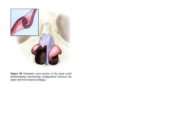
Note: Insert Picture, Upper and Lower Lateral Cartilage scrolls interlocking


Enhanced by Zemanta

Saturday, January 1, 2011

Viability of diced and crushed cartilage with different supportive sleeve material

Diced and crushed cartilage can be wrapped in different sleeve material. One of the main purposes for this is to camouflage irregular firm jagged edges of cartilage for a smoother cosmetic result especially in those with thin skin. Nose Revision Specialists have used the following materials as sleeves or fillers: Autogenous deep temporal fascia, Perichondrium, Surgicel (oxidized methylcellulose), dermal grafts, synthetic material and  esterified hyaluronic acid (HYAFF). A couple of study's have indicated the superiority of using deep temporal fascia or  esterified hyaluronic acid over surgicel (The Turkish delight method). I haven't come across any clinical experimental study's on Perichondrium  viability- reabsorption rate comparisons .  Dr. Eugene  A. Chu of John Hopkins University Dept. of Otolaryngology makes a recommendation to remove the perichondrium completely from the costal cartilage to limit warping, however Dr. Dean Toriumi, has claimed years of success with using perichondrium which will make the skin thicker and more importantly  help prevent graft visibility and deformity. Apparently it has given him a better long term outcome. Even though a few Surgeons have claimed success on long term follow up's with the Turkish delight method, most clinical studies indicate it is an inferior material to use, due to higher reabsorption rate of the contained cartilage.

According to Dr. Ozcan Cakmak and Dr. Fuat Buyuklu:

Some authors have stated that autogenous soft tissue grafts, such as dermal grafts3 or temporalis fascia grafts,5 are satisfactory for covering underlying dorsal irregularities. However, those materials have disadvantages of possible partial resorption and donor-site morbidity. Although some authors have suggested that temporalis fascia grafts are among the most reliable materials and are associated with reasonably low resorption rates,5, 8, 23 those grafts are difficult to manipulate because they are thin and slippery.23-24 Alloplastic materials, such as gelatin film,4 polyglactin 910 (Vicryl suture; Johnson & Johnson Gateway LLC, Piscataway, New Jersey),6 and Gore-Tex (W. L. Gore & Associates Inc, Newark, Delaware),7 have also been advocated for that purpose. However, absorbable synthetics do not last long, and of all graft materials, nonabsorbable synthetics are associated with the highest rates of infection and extrusion.2, 11 A soft-tissue filler, AlloDerm (LifeCell Corp, Branchburg, New Jersey), which is derived from cadaveric skin, is another material frequently used to achieve a smoother nasal dorsum. However, AlloDerm has the definite disadvantage of partial graft resorption, especially when it is positioned over the dorsum in patients with thin skin.

Although the maintenance of typical cartilage viability and a high graft survival rate have been reported with the use of this material, harvesting the temporalis fascia adversely affects the donor site, where either permanent or transient alopecia can develop after surgery.

Many authors have used bare crushed cartilage grafts to conceal dorsal irregularities and to achieve a smoother nasal surface.

http://archfaci.ama-assn.org/content/9/5/352.full

http://www.eclips.consult.com/eclips/article/Plastic-and-Aesthetic-Surgery/S1535-1513%2808%2970595-2

http://www.ncbi.nlm.nih.gov/pubmed/17690607

http://emedicine.medscape.com/article/881443-treatment

http://deantoriumi.com/faq1.asp

Saturday, December 25, 2010

Crushed vs Diced Cartilage: Similarities and Differences

Looking at some forums I noticed some people are using the words crushed and diced cartilage interchangeably as if they are same thing. Another important  issue i would like to address here is correlation between  the degree of crushed cartilage and how it effects the long term outcome (re absorption rate) of the surgery.

Crushing cartilage is procedure that takes pieces of cartilage and crushes it in a device called Cottle cartilage crusher and/or using a mallet. They can be crushed to varying degree's from slightly crushed to severely crushed. They are then inserted in the desired area of the nose using a medical tweezer.

Dicing cartilage is procedure where the cartilage is sliced using a straight edge razor blade into small fine pieces.  The cartilage is normally diced into <0.5mm squares, using two #11 blades avoiding, not being morselized or crushed. . Then it is placed in a syringe to be later injected in the desired area and molded/shaped accordingly. Usually diced cartilage  is wrapped in soft material preferably deep temporal fascia.

Both crushed and diced cartilage is used to smoothen out or camouflage nasal surface area's where cartilage is placed in the nose , like the dorsum, to conceal any irregularities. Both can be wrapped or combined with different material as well.

Crushed cartilage grafts can be used for the following purposes: (1) to cover the sharp edges of an irregular nasal framework after hump resection    (2) to serve as an underlying padding material to prevent skin adhesion   (3) to fill pit holes and, thus, mask irregularities   (4) as a filler to mask asymmetries and depressions on the side walls  (5) for tip grafting   (6) to camouflage the edges of solid onlay grafts  , (7) to supply minor dorsal augmentation for the correction of an overresected dorsum, and (8) to increase the thickness and natural color of the overlying skin where skin atrophy had occurred. I believe diced cartilage can be used in most of the above situations as well.

Crushed Cartilage Grafts for Concealing Irregularities in Rhinoplasty
  1. Ozcan Cakmak, MD;
  2. Fuat Buyuklu, MD

Our current clinical series confirmed our previous animal9 and human cell culture13 studies that the degree of crushing applied is important to the long-term clinical outcome of crushed cartilage grafts used in rhinoplasty.

The results showed a correlation between the degree of crushing applied and the resorption rate of the crushed graft, especially in grafts applied at the dorsum. The resorption rate was zero in slightly crushed grafts, 2.1% in moderately crushed grafts, and 13.1% in significantly crushed grafts. Our results show that slight or moderate crushing of the autogenous cartilage produces an outstanding graft material that is effective in concealing irregularities, filling defects, and creating a smoother surface, with excellent long-term clinical outcome and predictable esthetic result. We suggest that intact cartilage should be used to correct major deformities and that moderately crushed grafts should be used for smaller depressions to minimize resorption. The severely crushed form of cartilage should not be used as filler except to correct negligible depressions in atrophic skin.

The edges of solid onlay grafts might be softened by placing small pieces of moderately crushed grafts on or around the solid graft. The tiny pieces of moderately or significantly crushed grafts might be successfully used in final contouring at the conclusion of surgery. In patients with thin skin or in whom revision is required, a thin layer of moderately or significantly crushed cartilage would be the proper option as a padding material to prevent the adhesion of skin and to camouflage the sharp edges of the nasal skeleton that might be visible after edema has subsided.

From the above study,  one would presume that  thinner and smaller diced cartilage would also have higher resorption rates then thicker larger pieces, but I haven't seen any clinical studies to support or contradict that conclusion. 

http://archfaci.ama-assn.org/content/9/5/352.full

Wednesday, December 22, 2010

The Role of Diced Cartilage Grafts in Rhinoplasty

The fundamental technique for the use of diced cartilage in rhinoplasty has been known for over 50 years. One of the most impressive uses of diced cartilage is in cranioplasty, which demonstrates that the individual pieces coalesce into a semirigid graft over time. The term diced cartilage graft may refer to several different types of cartilage, methods of preparation, and methods of containment. In the present report, only autogenous cartilage derived from excised material, septum, or distant grafts is used. Containment refers to placement of the diced cartilage directly into a tight pocket for contour, layering of the cartilage on either side of rigid dorsal graft for blending, or placement of the cartilage in peripyriform pockets to advance the midface. The technique and benefits of diced cartilage grafts in rhinoplasty were reviewed.

A prospective study of more than 150 patients in 3 years found no evidence of absorption and no warping. Any problems thus far with the diced cartilage graft have been technical problems rather than problems with the graft material itself. One problem has been the visibility of radix grafts, particularly in patients with very active eyebrows. This problem is easily corrected by reduction with a pituitary rongeur or replacement with fascia alone. Dorsal grafts may have “edge show” cephalically, and caudally there may be inadequate grafting of the supratip region. This problem is easily corrected with the patient under local anesthesia by use of a pituitary rongeur. A minor depression may develop in the supratip area because the surgeon has initially undercorrected in pursuit of an immediate supratip break. This problem is corrected by keeping the graft truly full length rather than shortening it to get tip set off.
Conclusions: 
In using diced cartilage grafts in rhinoplasty, diced cartilage wrapped in fascia is simpler to use, quicker, and aesthetically superior to solid cartilage grafts, without risks of warping, malalignment, and K-wire extrusion.
 http://www.eclips.consult.com/eclips/article/Plastic-and-Aesthetic-Surgery/S1535-1513%2808%2970596-4

For the author,Rollin K. Daniel, MD; diced cartilage grafts have revolutionized dorsal grafts in rhinoplasty, replacing layered septal grafts, stacked conchal grafts, and carved costal cartilage grafts. He asserts that diced cartilage wrapped in fascia is simpler to use, quicker, and aesthetically superior to solid cartilage grafts, without risks of warping, malalignment and K-wire extrusion.

Diced cartilage grafts in rhinoplasty surgery: current techniques and applications. 

Dr.Rollin K. Daniel has used diced cartilage grafts in nasal surgery for more than 30 years. However, the number of cases and the variety of techniques have increased dramatically over the past 6 years.  

http://www.ncbi.nlm.nih.gov/pubmed/19050542


Autogenous Dorsal Reconstruction: Maximizing the Utility of Diced Cartilage and Fascia
Jay Calvert, M.D., F.A.C.S.1,2 and Kevin Brenner, M.D.2
 The problem of reconstructing the dorsum of the nose is complex and a source of frustration for both patients and surgeons. Dorsal deficiencies due to various etiologies and the need for dorsal contouring cause the plastic surgeon to look to time-honored techniques such as osseocartilaginous rib grafts while also searching for other options that may be less technically challenging and have the benefit of temporal success. Diced cartilage wrapped with deep temporal fascia is just such a method to achieve reliable dorsal reconstructions. The various ways to use diced cartilage and deep temporal fascia are discussed

The complications of using this technique are predictable and correctable. Because the cartilage is mobile for 10 to 14 days after placement, there can be defects that arise from poor management of the graft postoperatively. Edges are usually not visible, but they can be in a particularly thin-skinned patient. Overcorrection and undercorrection are probably the most common complications seen with this technique and must be managed accordingly. Malposition of the graft and mobility of the graft may also be seen in a rare number of cases. Absorption of the graft has not been seen in the longest of follow-ups (6 years).

In conclusion, the technique of diced cartilage with fascia (DC-F ) has been a useful method of dorsal reconstruction as a stand alone technique and in concert with other methods of building the dorsum. There are many permutations and surgical variations of the technique. The authors believe that proper preoperative analysis will help the surgeon to derive clear indications so that the correct graft variation is used with a clear purpose. There is no substitute for preoperative diagnosis and planning when using the DC-F graft. The technique is safe, easy to perform, has minimal morbidity, and is our favored method for addressing difficult problems in dorsal reconstruction.
Enhanced by Zemanta

Monday, December 20, 2010

Perichondrium vs Deep Temporal Fascia

When performing Augmentation or Revision nose surgery, soft tissue is needed. For instance it is used to cover cartilage used to build up area along the dorsum or tip or simply for augmenting an area, say's Dr. Paul Nassif. If doing Rib Harvesting, you can then use the Perichondrium which is soft tissue that lays on top of the rib instead of using temporal fascia. According to Dr. Nassif, it's a little more thicker, heavier, and firmer then Temporal fascia. He feels it's an excellent source of soft tissue. He also has a you tube video on how deep temporal fascia is harvested, but warning it is not for weak at heart. I assume he use's deep temporal fascia as a choice when not performing rib cartilage graft, to build up the radix or dorsum area's. This video  on harvesting Perichondrium.is less graphic, but still takes place in Operating Room.